

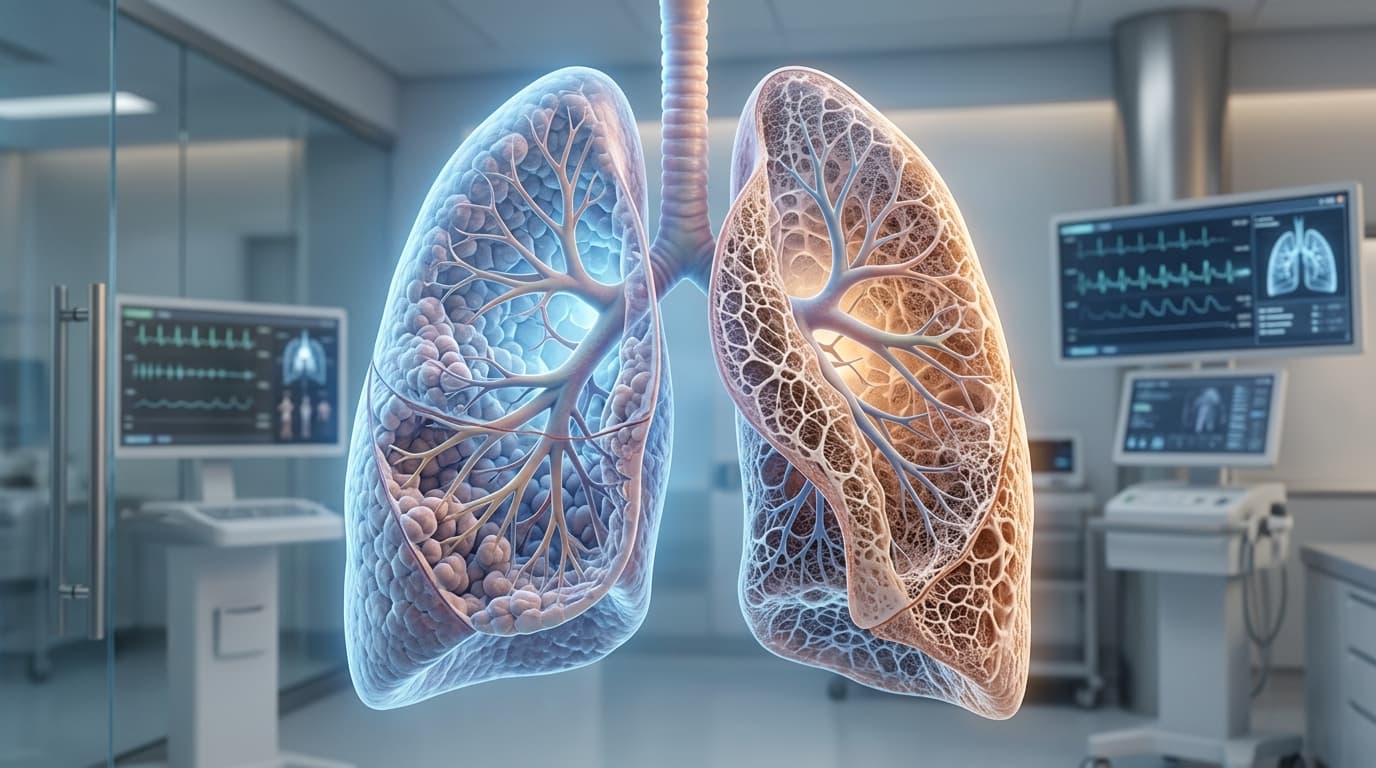
Pulmonary fibrosis is scarring of the lung tissue. Healthy lung is soft and elastic; it expands easily and lets oxygen pass efficiently into the blood. Scarred lung is stiff and thickened. It does not expand as well, and oxygen crosses it less easily.
The scarring itself does not reverse. That single fact shapes every clinical decision that follows — which is why the timing of diagnosis, and the timing of certain conversations, matters more in this condition than in almost any other respiratory disease.
What causes pulmonary fibrosis?
Fibrosis has many distinct causes, and identifying which one is present changes the treatment entirely:
- Hypersensitivity pneumonitis from inhaled triggers such as bird proteins or moulds
- Connective tissue and autoimmune diseases — rheumatoid arthritis, scleroderma, Sjögren's syndrome, and systemic sclerosis
- Occupational and environmental exposures — silica, asbestos, coal, and fine industrial dusts
- Medication-induced lung toxicity — including certain antiarrhythmics, chemotherapeutic agents, and immunosuppressants
- Post-infectious scarring — including long-term sequelae following severe viral pneumonia or ARDS
- Idiopathic pulmonary fibrosis (IPF) — where no underlying secondary cause is identified
Some forms of fibrosis are treatable at the source. Removing an antigen or controlling an autoimmune disease can halt progression. This is why an exhaustive diagnostic workup is not a formality — it decides whether the disease can be arrested or only slowed.
Idiopathic pulmonary fibrosis, specifically
Idiopathic pulmonary fibrosis (IPF) is the form where no specific cause can be found, and it is the most progressive condition in the interstitial lung disease spectrum. It tends to advance steadily rather than fluctuate.
Two clinical realities must be understood plainly:
- IPF is commonly diagnosed 12 to 24 months after initial symptoms begin. Early symptoms — a dry cough, breathlessness climbing stairs — are routinely attributed to ageing, body weight, past smoking, or lack of physical fitness.
- Without anti-fibrotic treatment, median survival from diagnosis is historically around three to four years. That is a population median, not an individual forecast, and contemporary therapy has improved this outlook. But it explains why a one-to-two-year diagnostic delay is so costly.
Persistent, gradually worsening breathlessness deserves systematic investigation, not reassurance. A high-resolution chest CT (HRCT) and lung function testing with DLCO answer the question quickly and reliably.
What medical treatment can and cannot do
Anti-fibrotic medicines have fundamentally altered the landscape of IPF management. They are prescribed to slow the rate of lung capacity decline and reduce the frequency of acute exacerbations.
Crucially, anti-fibrotics do not reverse existing scarring. Patients starting these medications sometimes anticipate feeling immediate symptomatic relief, and instead feel roughly unchanged while the underlying disease advances more slowly. That is the medication working as intended. Understanding this in advance prevents premature treatment discontinuation.
Alongside anti-fibrotic pharmaceuticals, comprehensive supportive care significantly improves quality of life:
- Pulmonary rehabilitation: Structured exercise and breathing retraining, representing one of the most effective non-pharmacological interventions for exercise tolerance.
- Management of secondary pulmonary hypertension: Screening for and treating elevated pressures in the pulmonary circulation.
- Timely vaccination: Comprehensive protection against influenza, pneumococcal disease, and respiratory viruses that trigger catastrophic declines.
- Nutritional preservation: Preventing muscle wasting and unintended weight loss that compromise future transplant candidacy.
- Gastroesophageal reflux management: Acid reflux is highly prevalent in fibrotic lung disease and micro-aspiration may accelerate disease progression.
- Ambulatory and resting oxygen therapy: Correcting hypoxemia to preserve organ function and relieve exertional fatigue.
When lung transplant becomes a question
For carefully selected patients with irreversible, advanced lung fibrosis, lung transplantation offers longer survival and restored functional independence.
Specific physiological indicators that should prompt an initial transplant evaluation include:
- A rising supplemental oxygen requirement during rest or minimal exertion
- Declining lung function parameters on serial testing, particularly a falling FVC or DLCO (>10% drop)
- Repeated hospital admissions for respiratory infection or acute exacerbations
- Progressive loss of physical stamina despite maximal medical therapy
When these markers appear, the question is not 'should we operate immediately?' The question is 'should this patient be evaluated by a dedicated transplant team?' Early assessment does not commit anyone to surgery — it creates a baseline and resolves treatable risk factors in advance.
Why timing decides the outcome
Timing is the factor most often missed, and delay can forfeit a patient's window of eligibility.
Referral should occur while a patient can still walk, eat independently, and maintain functional muscle mass. Patients evaluated at this ambulatory stage experience substantially better surgical outcomes than those referred in acute respiratory failure after prolonged intensive care.
By the time a patient has been ventilated or placed on emergency ECMO in an ICU for weeks, critical muscle deconditioning, secondary hospital-acquired infections, and multi-organ strain make major thoracic surgery exceptionally hazardous.
An early evaluation that concludes transplantation is not yet needed is a good clinical outcome: it establishes monitoring protocols so the team is prepared if disease kinetics accelerate.
What transplantation actually involves
Lung transplantation is major, complex surgery that demands complete transparency regarding benefits and demands.
Transplantation replaces a fatal, irreversible lung disease with a manageable chronic condition. It requires lifelong immunosuppressive medication to prevent allograft rejection, which introduces increased susceptibility to infections and potential renal impacts.
Regarding long-term outcomes: median survival following bilateral (double-lung) transplantation is approximately eight years and continues to improve with contemporary critical care and organ preservation techniques. For selected individuals facing end-stage respiratory failure, transplantation represents a transformative opportunity for renewed breath.
What to do if you have been diagnosed with lung scarring
If a chest scan has revealed pulmonary fibrosis or interstitial lung disease, take these six systematic steps:
- 1. Establish the precise underlying cause: Identifying an environmental antigen or autoimmune condition can halt progression.
- 2. Obtain baseline pulmonary function tests including DLCO: Serial trends over 3 to 6 months dictate disease trajectory.
- 3. Inquire whether anti-fibrotic medication is clinically indicated for your specific HRCT pattern.
- 4. Enroll in pulmonary rehabilitation early, while exercise capacity is preserved.
- 5. Protect nutrition and skeletal muscle mass proactively.
- 6. Request an early consultation with a transplant pulmonologist before urgent crisis arises.