

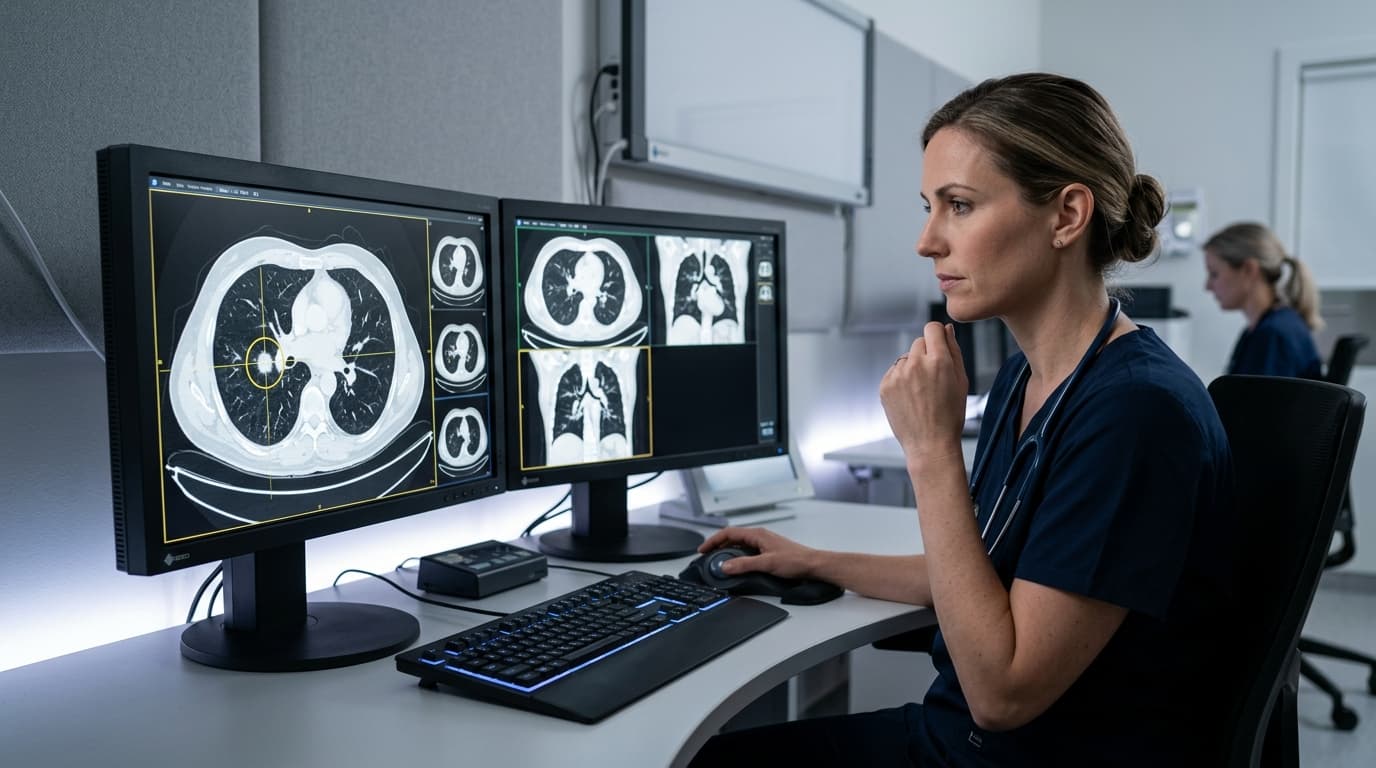
If you are reading this soon after seeing the words "pulmonary nodule" on a scan report, start here: most lung nodules are not cancer. The large majority turn out to be old infection, scarring, or a small benign growth that has been there for years and will stay unchanged for years more.
Nodules are found frequently, often by accident — during a scan done for a chest infection, a cardiac check-up, a pre-operative workup or a corporate health package. Modern CT scanners are detailed enough to pick up findings of a few millimetres that older scans never showed.
That said, "most are not cancer" is not the same as "ignore it." The purpose of what follows is to sort the ones that need attention from the ones that do not, without unnecessary surgery and without unnecessary anxiety.
What a lung nodule actually is
A pulmonary nodule is a small, roughly rounded area of increased density in the lung — under 3 cm across. Anything larger is called a mass and is assessed differently.
Reports describe them in several ways, and the wording matters:
- Solid nodule — uniformly dense
- Ground-glass nodule — hazy, less dense, sometimes significant even when small
- Part-solid — a mix of both, which warrants closer attention
- Calcified — contains calcium, which usually points strongly towards an old healed infection
Why old TB scars matter so much in India
This is the part that is often missed in international guidance, and it changes how nodules should be interpreted here.
Tuberculosis exposure is common across India. A very large number of people have had TB infection at some point, often without ever knowing. What it leaves behind is a small area of healed, frequently calcified scar tissue — and on a CT scan that appears as a nodule.
So a nodule with dense or central calcification, in someone from a high-prevalence region, is most often an old healed granuloma. It requires no treatment. It does not become cancer. Reacting to it with surgery would be a serious over-response.
Histoplasmosis and other fungal exposures leave similar marks, particularly along the Gangetic plain.
None of this means calcification is an automatic all-clear — the pattern of calcification matters, and a radiologist assesses it specifically. But it is a major reason why nodules in Indian patients should be interpreted by a specialist who sees this population regularly.
What size tells you
Size is the strongest single predictor of risk in solid nodules:
- Under 6 mm — risk is very low in people without major risk factors. Often no follow-up is needed at all, or a single check after a year.
- 6 to 8 mm — intermediate. Usually followed with a repeat CT at defined intervals to see whether it changes.
- Above 8 mm — warrants more active evaluation, which may include PET-CT or tissue sampling depending on the overall picture.
These are general principles, not a rule to apply to yourself. A 5 mm nodule in a 65-year-old with a 40-year smoking history is a different proposition from a 5 mm nodule in a 30-year-old non-smoker.
The single most useful thing you can do
Find your old scans.
A nodule that is identical on a CT from three years ago is, in almost all cases, benign. Something that has not changed in three years is not behaving like a cancer. That comparison can settle the question immediately, without a biopsy, without a PET scan and without surgery.
People routinely forget they had a CT during a COVID illness, before a knee operation, or in a health check. It is worth checking, and worth bringing the actual DICOM CD rather than only the report — the images allow direct measurement rather than relying on someone else's recorded number.
Always bring the actual DICOM CD with the scan images, not just the printed paper report. Specialist pulmonologists measure nodule dimensions and morphology directly.
What else affects the assessment
A specialist combines all of these factors rather than reacting to size alone:
- Age, since risk rises with it
- Smoking history, including how much and how long ago you stopped
- Previous cancer anywhere in the body
- Family history of lung cancer
- Occupational exposure to asbestos, silica, or radon
- Nodule location — upper lobe nodules carry slightly higher risk
- Nodule margins — smooth edges are reassuring, spiculated or irregular edges less so
- Growth rate, if serial scans exist
When is a biopsy needed — and which kind
If a nodule is suspicious enough to require tissue, the next question is how to obtain it with the least invasion:
- EBUS-TBNA — ultrasound-guided bronchoscopy, used particularly when enlarged lymph nodes in the centre of the chest need sampling. No incision, day-care procedure.
- CT-guided needle biopsy — generally better for nodules sitting near the outer edge of the lung.
- Navigational or radial EBUS bronchoscopy — for some peripheral nodules, reaching them through the airways.
- Surgical biopsy — appropriate in some situations, but it should generally not be the first step for an indeterminate nodule.
Being advised to remove part of a lung for a nodule that has not yet been characterised is worth a second look. In many cases the nodule can be diagnosed without surgery, and in some cases it turns out to require no intervention at all.
When to seek a second opinion
Reasonable circumstances to seek an expert second opinion include:
- You have been advised lung surgery for a nodule that has not been biopsied
- No one has compared the nodule against any previous scan
- The plan is unclear, or you have been told simply to "repeat in six months" with no explanation of what is being watched for
- The report mentions a part-solid or ground-glass nodule, which needs specific interpretation
- You want confirmation before committing to either surgery or years of surveillance
What to bring to the consultation
To make your consultation decisive and prevent repeated visits, bring:
- All CT scans on CD, not just reports — the images are what get measured
- Any older scans, even unrelated ones (prior health checks, abdominal or cardiac scans)
- Previous chest X-rays
- History of TB, TB treatment, or TB contact in the family
- Smoking history, including quantity and dates
- Occupational exposure history
- Details of any previous cancer
The realistic outcome
For most people the sequence is: the nodule is characterised, compared against prior imaging where available, and either dismissed or placed under a defined surveillance schedule. A minority need tissue. A smaller minority need surgery.
The interval between finding a nodule and understanding it is uncomfortable. It is usually short, and it usually ends with reassurance.