

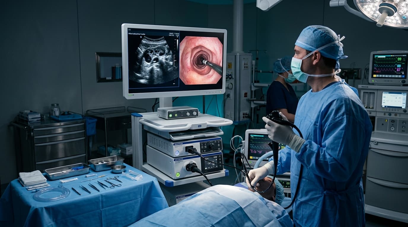
If you have been told you need an EBUS, you have probably already read what it is. What most people actually want to know is different: will it hurt, will I be awake, how long does it take, and when do I get the answer.
This guide covers those questions directly and transparently.
What it is, briefly
EBUS-TBNA stands for endobronchial ultrasound with transbronchial needle aspiration. A thin flexible tube with an ultrasound probe at its tip is passed through the mouth into the main airways. The ultrasound looks through the airway wall at the lymph nodes and structures sitting behind it, and a fine needle is passed through the wall to take a sample under direct ultrasound vision.
The important part is what it replaces. Obtaining tissue from the centre of the chest used to require mediastinoscopy — a surgical procedure with a neck incision, general anaesthesia and a hospital stay. EBUS achieves much of the same diagnostic information through the natural airway, with no incision at all.
Why it might have been recommended
Common reasons include enlarged lymph nodes in the centre of the chest seen on a CT scan, a suspected lung cancer requiring staging, suspected sarcoidosis or tuberculosis, or a mass sitting adjacent to a major airway.
It is a diagnostic procedure. Its purpose is to answer a question accurately, and often the answer is reassuring — a great many EBUS procedures done for "suspicious" nodes return sarcoidosis or tuberculosis rather than cancer.
Before the procedure
To ensure safety and procedure accuracy, you will usually be asked to:
- Fast for around six hours beforehand — no food, and clear fluids only up to a specified time.
- Have blood tests, typically including clotting studies.
- Discuss blood thinners. Aspirin, clopidogrel, warfarin and newer anticoagulants may need pausing for a set number of days. Never stop these on your own — the instruction has to come from the team, weighing bleeding risk against the reason you are taking them.
- Mention diabetes medication, inhalers, allergies, loose teeth or dentures, and any previous reaction to sedation.
- Arrange someone to accompany you home. You will have had sedation and cannot drive.
Never stop blood thinners or antiplatelet drugs on your own. The medical team must give explicit instructions based on procedure risk versus cardiac protection.
What happens during the procedure
You will not be fully awake and aware of it. The procedure is done under conscious sedation — medication through a drip that leaves you drowsy and comfortable, with little or no memory of it afterwards. Some centres use deeper sedation or general anaesthesia depending on the case. This will be explained beforehand.
Your throat is numbed with a local anaesthetic spray, which tastes bitter and makes the throat feel thick. Oxygen levels, heart rate and blood pressure are monitored throughout.
The scope is passed through the mouth, past the vocal cords and into the airways. It does not block your breathing — this is the concern most people have, and it is worth stating plainly. The scope is far narrower than the airway it sits in. You breathe normally around it throughout.
The ultrasound identifies the target. A needle is passed through the airway wall into the lymph node and samples are taken, usually several passes from each site. There are no pain-sensing nerves inside the airways in the way there are on skin, so this is not felt as sharp pain.
Typical duration is 30 to 45 minutes, though it varies with how many sites need sampling.
Some centres use ROSE — rapid on-site evaluation — where a pathologist examines a smear immediately to confirm adequate material has been obtained before the procedure ends. It reduces the chance of needing a repeat.
Afterwards and recovery
Recovery is rapid and straightforward for the vast majority of patients:
- You rest in recovery while the sedation wears off, usually one to two hours.
- No eating or drinking until the throat numbness has worn off, typically around two hours. Swallowing before sensation returns risks food going the wrong way.
- A sore throat and hoarse voice for a day or two are normal.
- Coughing, and some blood-streaking in the sputum for a day, is expected and not a cause for alarm.
- Most people are discharged the same day, generally within three to four hours.
- You may feel tired for the rest of the day. Most people return to normal activity the next day.
Contact the hospital or go to emergency immediately if you develop significant fresh bleeding, worsening breathlessness, severe chest pain or fever.
Risks, stated honestly
EBUS is a low-risk procedure, but it is an invasive procedure, and no one should consent to it without knowing the actual risks:
- Minor bleeding at the puncture site — common, almost always self-limiting
- Sore throat and cough — very common, short-lived
- Sedation-related effects such as a drop in blood pressure or oxygen — monitored for and managed
- Infection, including rarely a mediastinal infection — uncommon
- Pneumothorax (collapsed lung) — uncommon with EBUS, because the needle passes into structures adjacent to the airway rather than through lung tissue as a CT-guided biopsy does
- Non-diagnostic sample, requiring a repeat or different approach
Ask your team for their own complication rates. A high-volume interventional pulmonology operator should be able to answer that question directly.
When do results come back?
Turnaround time depends on what is being investigated:
- Routine cytology and histology — usually within 48 hours to a few days
- TB culture — several weeks, though rapid molecular tests (such as GeneXpert) return within a day or two
- Molecular and genetic testing on cancer samples — one to three weeks, since these guide targeted treatment and take longer
How it compares to the alternatives
The choice depends on where the target sits. Location, not preference, determines the approach:
- CT-guided needle biopsy — better for nodules at the outer edge of the lung, but carries a meaningfully higher pneumothorax risk and cannot reach central lymph nodes.
- Mediastinoscopy — surgical, requires general anaesthesia and a neck incision, with a hospital stay. Still has a role in specific situations, but EBUS has replaced it for many indications.
- Surgical lung biopsy — more invasive, appropriate in some circumstances but rarely the right first step for central lymph nodes.
Questions worth asking beforehand
Key questions to ask your interventional pulmonologist before the procedure:
- 1. What exactly are you sampling, and why that site?
- 2. What sedation will I have?
- 3. Will ROSE (rapid on-site evaluation) be used?
- 4. How many of these do you perform, and how often?
- 5. When will I get results, and who will explain them?
- 6. What happens if the sample isn't diagnostic?