

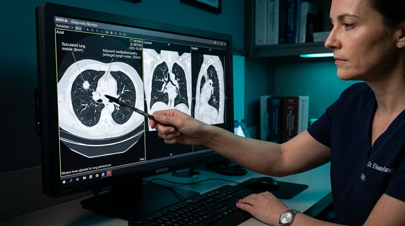
In London or Boston, a doctor who sees an enlarged lymph node in the centre of a patient's chest has one dominant thought. Tuberculosis is uncommon there. Sarcoidosis is possible. But statistically, an enlarged mediastinal node in an adult is most likely to be cancer, and the entire diagnostic pathway is built on that assumption.
In India, that assumption is wrong often enough to be dangerous.
Tuberculosis is endemic here. A very large number of Indians have been infected at some point, most without ever knowing. TB affects lymph nodes readily. So when a scan shows an enlarged node in the chest, or a single round shadow in the lung, the honest answer is that we do not yet know what it is.
In my own practice, looking at patients referred with a solitary lung nodule, roughly one in three has turned out to be cancer. The remaining two-thirds were tuberculosis or another infection.
That ratio is the entire reason this article exists. It means a doctor in India cannot reason their way to a diagnosis from a scan. Neither guess is safe. The tissue has to be obtained.
What a solitary lung nodule actually is
A solitary pulmonary nodule is a single, roughly round area of increased density in the lung, usually under 3 centimetres across, with normal lung around it. It is found constantly — during a scan done for a chest infection, before surgery, in a corporate health check, or while investigating something else entirely.
Modern CT scanners are sensitive enough to detect lesions of a few millimetres that older imaging never showed. Most of what they find is harmless.
In a Western population, "most of what they find is harmless" mostly means benign growths and old scarring from fungal infection. In India, it overwhelmingly means healed tuberculosis.
TB leaves behind small areas of scar tissue, often calcified, that persist for life. A person who had a subclinical TB infection at nineteen may carry that nodule at sixty and never know why it is there. On a CT scan, it can look very similar to something that needs urgent attention.
The two ways this goes wrong
Mistake one: assuming it is TB
This is by far the more common error in India, and it is entirely understandable. Anti-tuberculosis treatment is available, affordable and familiar. A patient with a cough, weight loss and a shadow on the chest X-ray fits the TB picture perfectly. Treatment is often started without microbiological confirmation.
Most of the time, that instinct is correct and the patient recovers. But when it is wrong, the cost is measured in months. Indian epidemiological work has found that a meaningful proportion of lung cancer patients — in one series, around 14 per cent — had been incorrectly started on anti-TB treatment, delaying their cancer diagnosis substantially.
Lung cancer does not pause during a six-month course of anti-TB drugs.
The practical rule: if anti-TB treatment was started without a positive sputum test, a positive GeneXpert, or a tissue diagnosis — and there is no clear improvement after four to six weeks — the diagnosis needs revisiting. Not at the end of the course. At six weeks.
Mistake two: assuming it is cancer
The opposite error is less discussed but also real, and it tends to happen at well-equipped centres.
A patient is told a nodule is suspicious. They are frightened. Sometimes they are advised surgery to remove part of the lung before any tissue has been obtained. Sometimes they travel, spend heavily, and undergo procedures for what turns out to be an old tuberculous scar that would never have caused them harm.
Given that roughly two-thirds of the nodules I see turn out not to be cancer, treating every suspicious shadow as malignant would mean subjecting a great many people to unnecessary surgery, cost and fear.
Being advised lung surgery for a lesion that has not been biopsied is worth a second opinion. In many cases the nodule can be characterised without an operation, and in some it needs no intervention at all.
And sometimes it is both
This is the complication that makes Indian practice genuinely difficult.
Tuberculosis and lung cancer can coexist in the same patient. A single-centre prospective Indian study reported coexistence in around 16 per cent of the patients studied, and outcomes were worse than with either condition alone.
The clinical trap is obvious once stated. A patient is sampled, TB is found, treatment starts, everyone is satisfied — and the cancer sitting alongside it is missed for months. Published case reports describe exactly this: patients with confirmed tuberculosis whose malignancy was only identified because someone sampled further.
Finding one diagnosis is not the same as excluding the other. Thorough multi-station sampling protects against this trap.
What actually settles the question
Comparing with older scans: The cheapest and often most decisive step. A nodule that appears unchanged on a CT from three years ago is, in almost all cases, benign — cancer does not stay the same size for years. It is worth looking, and worth bringing the actual DICOM CD rather than only the report, so the lesion can be measured directly.
What the scan itself suggests: Dense or central calcification points strongly towards old healed infection. Smooth margins are reassuring; spiculated, irregular margins less so. Upper-lobe location, cavitation patterns, surrounding tree-in-bud changes and adjacent nodes all contribute. But imaging shifts probability — it does not settle the question.
Tissue settles the question: This is the step that gets skipped, and it is the one that matters.
- EBUS-TBNA: For enlarged lymph nodes in the centre of the chest, EBUS-TBNA is the tool of choice. Done under conscious sedation, usually 30 to 45 minutes, with no incision, and home the same day.
- CT-guided needle biopsy: For nodules sitting at the outer edge of the lung, CT-guided biopsy is often better suited.
- Navigational or radial bronchoscopy: Peripheral lesions can also be reached through the airways with thin or radial ultrasound scopes.
Why EBUS matters so much in an Indian setting specifically
Four decisive factors make EBUS transformative in India:
- One sample answers both questions: The same material can be sent for cytology/histology to look for malignancy, and simultaneously for TB culture and rapid molecular testing such as GeneXpert. Cancer and TB are assessed in parallel rather than in sequence.
- Multiple stations can be sampled in one sitting: Reducing the chance of finding one and missing coexistent pathology.
- It avoids surgery for a benign diagnosis: Given that most of these nodules are not cancer, a minimally invasive route spares people operations they never needed.
- It provides enough tissue for molecular testing: Vital if the result is adenocarcinoma, where EGFR mutations are 2 to 3 times more frequent in India than in the West.
What to ask your doctor
If you have been told you have a nodule or an enlarged lymph node in the chest:
- 1. Has anyone compared this with an older scan? (If old imaging exists, this should happen before anything else)
- 2. Is there microbiological or tissue confirmation, or is this a presumptive diagnosis?
- 3. If I am being started on anti-TB treatment, when will we reassess if I do not improve?
- 4. Can this be sampled without surgery? (In most cases involving central nodes, it can)
- 5. If tissue is taken, will it be tested for both TB and malignancy?
- 6. If it is cancer, will molecular testing be done?
What to bring to the consultation
Prepare these materials for your specialist appointment:
- All CT scans on CD, not just the printed reports — the actual images are what get measured
- Any older scans, even unrelated ones (prior health packages or surgeries)
- Any history of TB, TB treatment, or TB in the household
- Previous chest X-rays
- Smoking history, including bidi use, and occupational dust or smoke exposure
- Details of any previous cancer or systemic health conditions
The point, stated plainly
A nodule on a scan is a question. In a country where tuberculosis is endemic, it is a genuinely open question — in my experience, cancer roughly a third of the time and infection the rest.
Guessing wrong in one direction costs months of a cancer patient's life. Guessing wrong in the other costs a healthy person part of a lung.
Neither is necessary. The answer is usually obtainable in a day-care procedure with no incision, and the great majority of people who go through it receive reassuring news.